Services
- Abdominal x-ray
- Abdominal Ultrasound
- Bone X-rays
- Cardiac MRI
- Carotid Doppler
- Chest X-Ray
- CT SCAN
- Echocardiography
- Electrocardiography (ECG)
- General Ultrasound Imaging
- General Doppler Imaging
- Magnetic Resonance Imagining
- Prostatic Ultrasound
- Scrotal Ultrasound
- Ultrasound of the Thyroid
- Venous Doppler
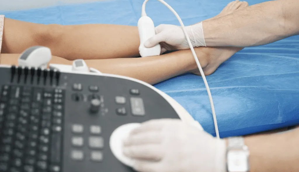
Professional medical service of Venous Doppler
Ultrasound imaging, also known as ultrasound scanning or sonography, is a safe and painless diagnostic technique that uses high-frequency sound waves to produce images of the body’s internal structures. A small probe and ultrasound gel are used to transmit sound waves, which create real-time images of internal organs and blood flow. Unlike X-rays, ultrasound does not use ionizing radiation.
Types of Ultrasound Imaging
- Conventional Ultrasound: Produces thin, flat images of the body.
- Doppler Ultrasound: Evaluates blood flow through major arteries and veins in areas like the abdomen, arms, legs, and neck.
Why is a Venous Ultrasound Performed?
Venous ultrasound primarily helps identify blood clots, such as deep vein thrombosis (DVT), which may lead to pulmonary embolism if untreated. Other Uses:
- Determine the cause of leg swelling due to conditions like varicose veins.
- Guide the placement of needles or catheters into veins.
- Map veins for surgical procedures like bypass grafting.
- Assess blood vessel grafts used for dialysis to identify narrowing or blockages.
Doppler Imaging Benefits:
- Detect blood clots, vessel narrowing, and abnormal blood flow.
- Evaluate tumors or malformations in vessels.
- Assess increased blood flow as a potential sign of infection.
Preparation Guidelines
- Clothing: Wear loose, comfortable clothing and remove jewelry near the examination area.
- Fasting: Required only for abdominal vein examinations (6-8 hours before).
- For most venous ultrasounds, no special preparation is needed.