Services
- Abdominal x-ray
- Abdominal Ultrasound
- Bone X-rays
- Cardiac MRI
- Carotid Doppler
- Chest X-Ray
- CT SCAN
- Echocardiography
- Electrocardiography (ECG)
- General Ultrasound Imaging
- General Doppler Imaging
- Magnetic Resonance Imagining
- Prostatic Ultrasound
- Scrotal Ultrasound
- Ultrasound of the Thyroid
- Venous Doppler
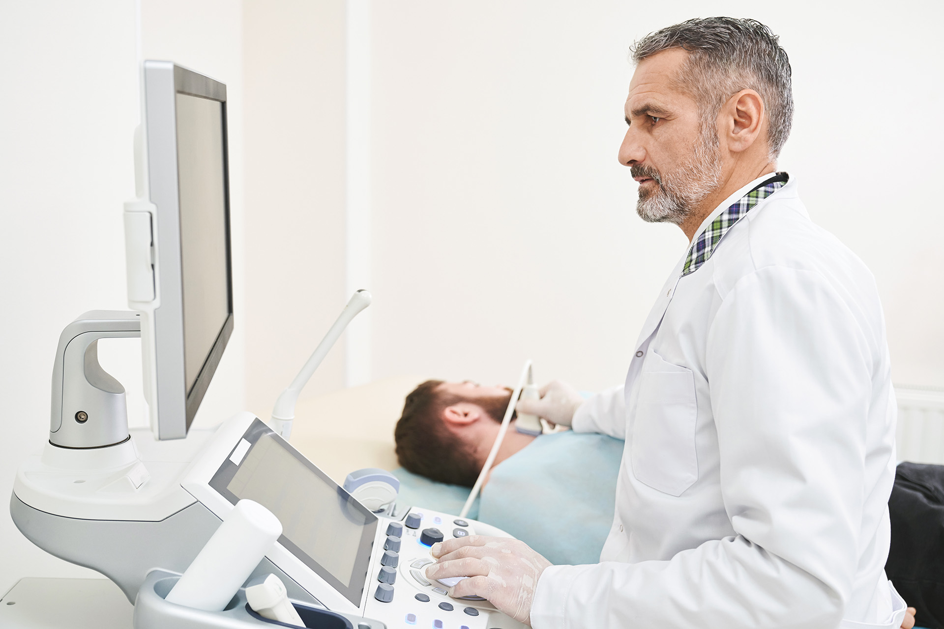
Professional medical service of Ultrasound of the Thyroid
Ultrasound imaging, also known as ultrasound scanning or sonography, uses a small probe and a special gel to expose the body to high-frequency sound waves. It is a safe, painless technique that creates images of the body’s interior using sound waves without involving ionizing radiation (as in X-rays). Because ultrasound captures real-time images, it can display the structure and movement of internal organs, as well as blood flow within blood vessels
Conventional ultrasound presents images as thin, flat sections of the body.
Doppler ultrasound is a specialized technique used to evaluate blood flow through vessels, including major arteries and veins in the abdomen, arms, legs, and neck
Why should you consider this procedure?
Ultrasound of the thyroid is typically performed to:
- Determine if a lump in the neck originates from the thyroid or an adjacent structure.
- Analyze the characteristics of thyroid nodules to identify whether they are benign or have features requiring a biopsy. Ultrasound-guided fine needle aspiration can enhance biopsy accuracy if needed.
- Detect additional nodules in patients with one or more nodules identified during a physical exam.
- Monitor significant growth of thyroid nodules over time.
Ultrasound’s real-time imaging capabilities make it valuable for guiding procedures, such as needle biopsies, to extract tissue samples for laboratory testing. It can also assist in the safe and precise insertion of catheters or drainage devices.
Preparation Guidelines
- Wear comfortable, loose-fitting clothing.
- You may need to remove clothing and jewelry from the area to be examined.
- For children, it is important to minimize movement, as ultrasound examinations are sensitive to motion. Preparing the child by explaining the procedure beforehand can help ensure a smoother process.