Services
- Abdominal x-ray
- Abdominal Ultrasound
- Bone X-rays
- Cardiac MRI
- Carotid Doppler
- Chest X-Ray
- CT SCAN
- Echocardiography
- Electrocardiography (ECG)
- General Ultrasound Imaging
- General Doppler Imaging
- Magnetic Resonance Imagining
- Prostatic Ultrasound
- Scrotal Ultrasound
- Ultrasound of the Thyroid
- Venous Doppler
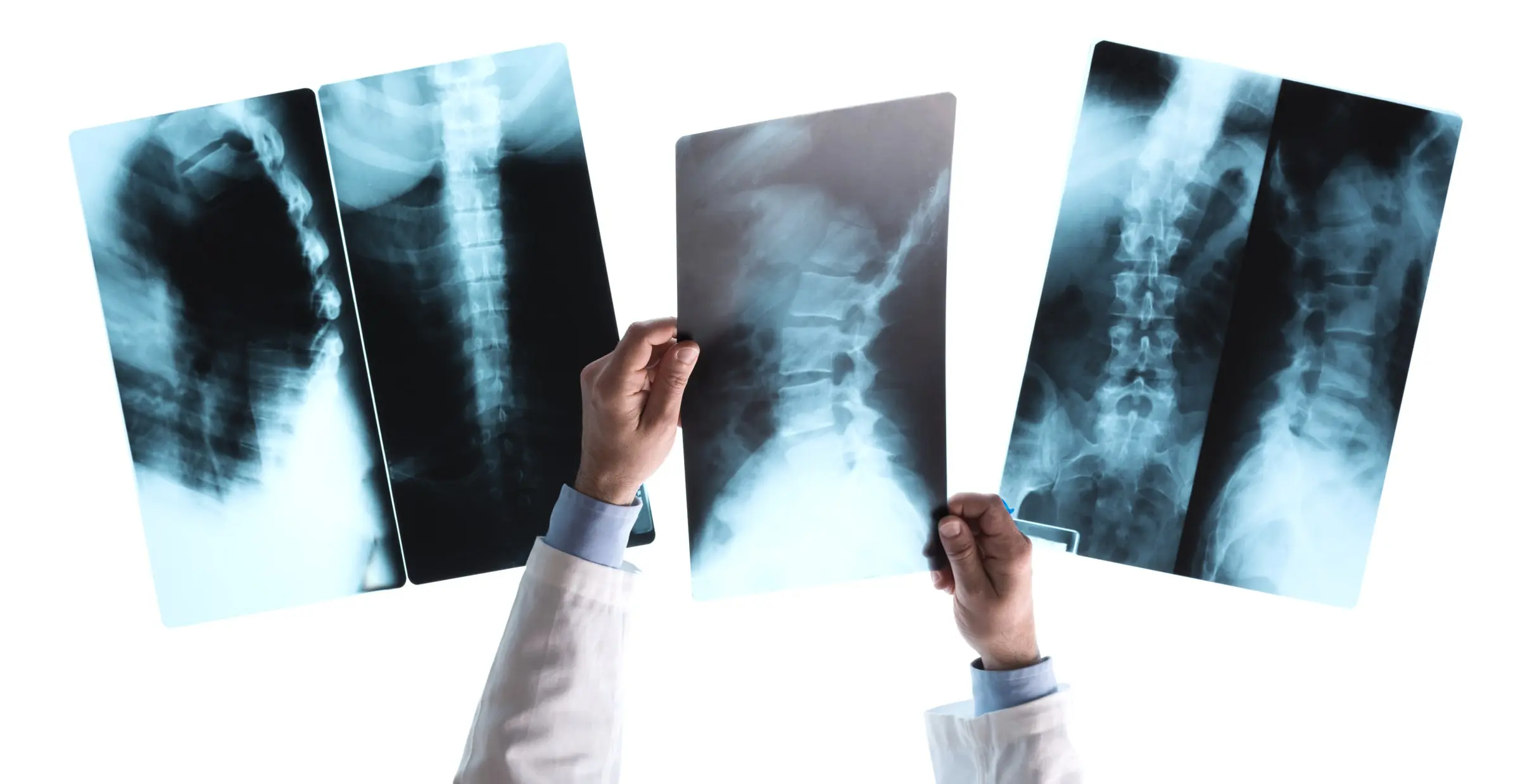
Professional medical service of Bone X-rays
An X-ray, or radiograph, is a noninvasive diagnostic tool used to help physicians diagnose and treat various medical conditions. It works by exposing a part of the body to a small dose of ionizing radiation, producing images of internal structures. X-rays are the oldest and most widely used form of medical imaging.
Uses of Bone X-Rays
Bone X-rays are commonly performed to:
- Diagnose fractures or joint dislocations.
- Verify proper alignment and stabilization of bones after fracture treatment.
- Guide orthopedic procedures, such as spine repair, joint replacements, and fracture reductions.
- Detect injuries, infections, arthritis, abnormal bone growths, and metabolic bone changes.
- Assist in identifying and diagnosing bone cancer.
- Locate foreign objects in soft tissues near or within bones.
Preparation Guidelines
- Most X-ray exams require no special preparation.
- You may be asked to remove some or all clothing during the exam.
- Remove any jewelry, removable dental appliances, eyeglasses, and other metal objects or clothing that might interfere with the imaging process.
- Women should always inform their physician and X-ray technologist if there is any possibility of pregnancy.