Services
X-ray unit
Fluoroscopy Imaging
Fluoroscopy is a medical imaging procedure that uses multiple pulses of X-rays to capture real-time moving images of tissues and organs inside the body. This technique is used by healthcare providers to monitor and diagnose certain medical conditions, as well as to guide various medical procedures.
What is the difference between fluoroscopy and traditional X-ray imaging?
Traditional X-rays produce still images of internal structures—similar to a photograph—while fluoroscopy functions more like a video, offering continuous moving images of internal organs and tissues through a series of radiation pulses.
Uses of Fluoroscopy
Fluoroscopy is primarily used for two purposes:
- Diagnostic Evaluation – To assess and monitor specific medical conditions.
- Interventional Guidance – To assist in guiding medical procedures such as surgeries, catheter insertions, and interventional radiology procedures.
How Does Fluoroscopy Work?
The procedure involves exposure to a low dose of ionizing radiation to produce clear images of internal structures, making it a safe and effective tool widely used in diagnostic imaging.
Preparation and Procedure
A fluoroscopy exam may involve the following steps:
- You may be asked to remove any clothing or jewelry that could interfere with the procedure. A medical gown will be provided if needed.
- In some cases, a contrast agent may be used to enhance the visibility of certain organs and tissues. The contrast may be administered in one of the following ways:
- Orally, by drinking a liquid containing the dye.
- Intravenously, through an IV catheter.
- Rectally, via an enema if the lower gastrointestinal tract is being examined.
- You may also be instructed to hold your breath briefly to obtain clearer images during the scan.
- You will then be positioned on an imaging table. Depending on the type of exam, you may be asked to change your body position or move specific body parts.
Why Choose Fluoroscopy?
Fluoroscopy is an ideal imaging option for visualizing organ motion in real time, making it highly effective in enhancing diagnostic accuracy and supporting interventional medical procedures.
If you need more information about the procedure or specific preparation instructions, please don’t hesitate to contact the RadCare team.
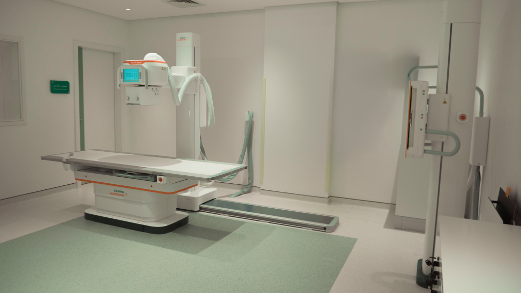
Fluoroscopy with Luminos dRF Max by Siemens – Al-Rabwa Branch
Key Features:
- 2-in-1 Solution: Combines fluoroscopy and digital radiography in one system, enhancing workflow efficiency.
- High Imaging Quality: Equipped with DiamondView Max image enhancement technology, delivering superior image quality with minimal radiation dose for faster, more confident diagnosis.
- Enhanced Safety: Intelligent design facilitates patient transfer and minimizes injury risks, especially for patients with special needs (table height as low as 48 cm).
- Ease of Use: Features smart control with the syngo FLC interface and SmartTouch joystick, plus automated smart movement via SmartMove.
- Radiation Control: Includes applications such as CAREPROFILE and CAREPOSITION to reduce unnecessary radiation exposure.
- Supports Digital Subtraction Angiography (DSA) and SmartOrtho for imaging long bones and the spine on a single film.
General X-ray with Siemens MULTIX Impact – Al-Namudhajiyah and Al-Nafal Branches
The Siemens MULTIX Impact is a digital radiography system designed to deliver reliable performance and high-quality imaging for fast, accurate patient examinations.
Key Features:
1. Flexible and Comfortable Design:
- Allows operators to remain close to patients during exams, reducing errors due to patient movement and improving overall comfort.
- The table offers lateral movement, making it easier to center different body parts under the X-ray beam.
- The table can be lowered to as little as 490 mm, facilitating access for elderly patients and children.
2. Smart Imaging with myExam Companion:
- Provides visual guidance and reduces human error, improving diagnostic accuracy and workflow efficiency.
3. Enhanced Patient Experience:
- Tube-integrated display: Enables staff to stay with the patient longer during the scan.
- Adjustable table height: Ranges from 51.5 cm to 90 cm, allowing easy access for elderly patients and those with special needs.
- Positioning guide: Helps improve patient alignment, minimizing the need for repeat scans and reducing radiation exposure.
4. Reliability and Speed:
- Delivers fast, standardized results.
- High-quality components ensure system durability and minimize downtime, enabling timely delivery of diagnostic results.