Services
Women’s Health Unit – Al-Namudhajiyah Branch
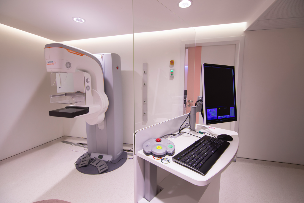
Mammography Department
Radcare offers the latest mammography technologies, considered the most effective method for early routine screening for breast cancer. Mammography is distinguished by its ability to detect early signs of cancer before any symptoms or noticeable changes appear to the woman or her physician.
How is a mammogram performed?
- The woman stands in front of the mammography machine, and one breast is gently placed on a plastic plate. Another plate is lowered from above to compress the breast and hold it in place, which helps produce a clear image. The compression lasts only a few seconds and does not cause harm.
- The same steps are repeated with the other breast. Afterward, the plates are tilted to capture side views of both breasts.
What are the benefits of this exam?
- Mammography is used as an early detection tool for breast cancer in women. It can also be used to detect and diagnose breast conditions when symptoms are present, such as:
- A lump
- Pain
- Nipple discharge
What preparations are needed?
- It is advised to perform the exam one week after your menstrual period to reduce breast sensitivity.
- Inform your doctor or technician if there is any chance you may be pregnant.
- Avoid using deodorant, powder, or similar products under the arms or on the chest area on the day of the exam.
- Inform the doctor or technician of any breast-related symptoms.
- Please bring previous mammograms and reports for comparison during the exam.
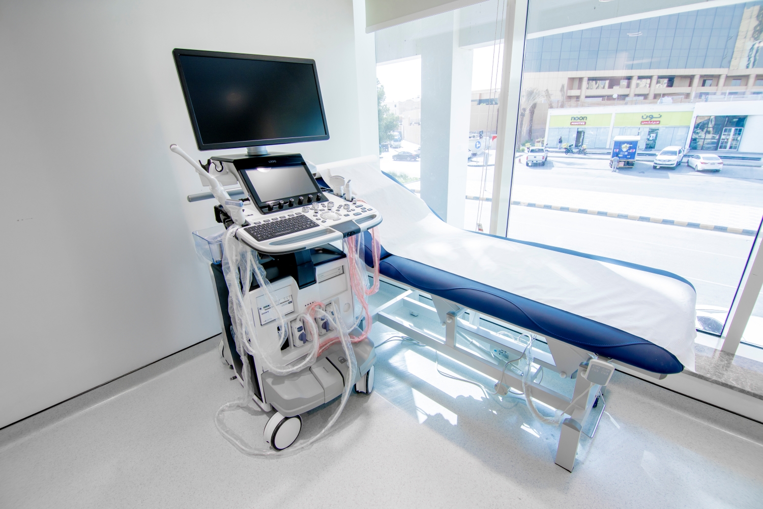
Ultrasound Department
GE Voluson E10 Women’s Health Ultrasound
At Radcare, we provide ultrasound services performed by skilled medical and technical staff using modern, high-precision equipment.
Ultrasound is a procedure that uses high-frequency sound waves to create visual images of the inside of the body. These sound waves reflect off tissues and are converted into images, helping visualize internal structures, movement of organs, and blood flow through vessels.
Traditional ultrasound provides flat, thin slices of the body. However, with technological advancements, sound wave data can now be used to create 3D ultrasound images, and even 4D ultrasound, which shows real-time movement in 3D.
This test is safe, painless, and does not use ionizing radiation, unlike X-rays.
Ultrasound Uses:
Breast Exam
- Guided tissue sampling (biopsy), using a needle to collect cells from abnormal areas for lab testing.
- Breast imaging and biopsy guidance in suspected breast cancer cases.
4D Ultrasound
4D ultrasound provides real-time imaging of fetal development and offers valuable insights into the baby’s condition before birth. Common benefits include:
- Fetal face: Helps detect congenital anomalies such as cleft lip.
- Stomach and abdomen: When the baby swallows amniotic fluid, the stomach appears as black bubbles. The scan can confirm the abdomen is covering internal organs well.
- Spine: Assesses spinal alignment and skin coverage to rule out abnormalities.
- Also useful for evaluating major arteries and veins that transport blood from the heart to various organs.
Additional Ultrasound Applications:
Ultrasound is useful for examining many internal organs, including:
- Heart and blood vessels (including abdominal aorta and major branches)
- Kidneys, pancreas, spleen, gallbladder, and liver
- Bladder
- Uterus, ovaries, and fetus in pregnant women
- Eyes
- Thyroid and parathyroid glands
- Infant brain
- Infant hips
Preparation Instructions:
- Wear comfortable, loose-fitting clothing.
- Remove any clothing, jewelry, or accessories from the area being examined.
- Other preparations depend on the type of scan:
- For some exams, you may be asked to fast for up to 12 hours beforehand.
- For pelvic or abdominal scans, you may be instructed to drink up to 6 cups of water two hours before the scan and avoid urinating to ensure the bladder is full.
For Children:
- It is advised to calm the child before the scan.
- Ultrasound is highly sensitive to movement, which may slow down or prevent the exam if the child moves excessively or cries.
Bone Densitometry Department
At Radcare, we use advanced bone densitometry devices that meet the highest standards of accuracy and quality.
This test determines whether you have osteoporosis.
Why should you get a DEXA scan (Bone Density Test)?
There are several reasons to consider a DEXA scan, including:
- Monitoring changes in bone health over time
- Assessing the effectiveness of osteoporosis treatment
- Estimating the risk of future fractures
- Confirming an osteoporosis diagnosis
People with a history of osteoporosis or frequent fractures should undergo the test regularly.
Frequently Asked Questions
Can I have a bone density test while pregnant?
- No. It is not recommended to perform a DEXA scan during pregnancy due to radiation exposure that may harm the fetus. You should inform your doctor if there is a possibility of pregnancy before the test.
Women’s Health Packages

Women’s Health Package
Description: A comprehensive program dedicated to women’s health care
Target Audience: Women of all ages
- Follicle Stimulating Hormone (FSH)
- Luteinizing Hormone (LH)
- Prolactin
- Vitamin D
- Thyroid Stimulating Hormone (TSH)
- Complete Blood Count (CBC)
- Estradiol
- Pelvic Ultrasound (Ovaries)
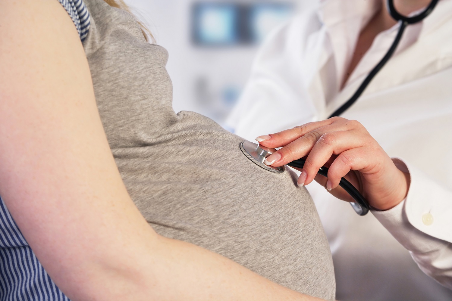
Pregnancy Follow-up Package
Description: A comprehensive program for maternal care during all pregnancy stages
Target Audience: Women at any stage of pregnancy
- Ferritin (Iron Stores)
- Complete Blood Count (CBC)
- Blood Sugar Test
- Vitamin D
- Calcium
- Ultrasound
Additional 4D Ultrasound available once during pregnancy for only 300 SAR
Check & Reassure Package
Description:A screening package for checking and reassuring women’s health
Target Audience: Women of all ages
- Breast Tumor Markers
- Mammogram