Services
Ultrasound Imaging Unit (Sonar & Doppler)
Sonar Department
Ultrasound (sonar) is a non-invasive and painless diagnostic method that can capture static or moving images of internal organs. This imaging technique relies on high-frequency sound waves.
What are the steps of undergoing an ultrasound scan?
The scan usually takes no longer than 30 minutes, depending on the area being examined.
- The scan usually takes no longer than 30 minutes, depending on the area being examined.
- You may be asked to undress and wear a gown, then lie on an exam table with the target area exposed.
- A technician will apply a gel to the skin over the area to be scanned. This gel prevents friction and enhances the transmission of sound waves.
- The doctor will move the transducer over the body to capture clear images.
- You might be asked to adjust your position slightly to allow more accurate imaging.
- The technician will wipe off any remaining gel after the exam.
- You can return to your normal daily activities immediately.
Doppler Department
Doppler ultrasound is a non-invasive imaging test used to evaluate blood flow through vessels.
It is a safer and simpler alternative to other vascular imaging methods because it does not require the use of contrast dye or exposure to X-rays.
It works by bouncing high-frequency sound waves off red blood cells in the bloodstream.
Uses of Doppler Ultrasound:
Detects damaged or narrowed arteries.
Monitors the impact of treatments on blood vessels.
Essential in detecting deep vein thrombosis (DVT)—commonly in the legs—which could lead to serious complications like pulmonary embolism.
Also used to monitor blood flow after surgeries.
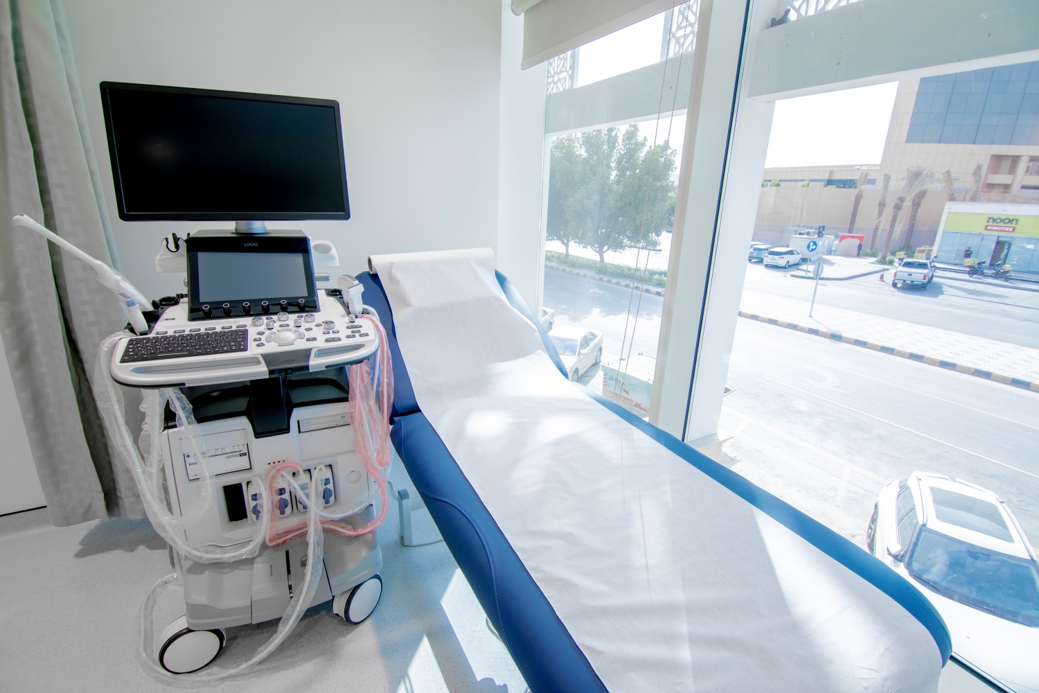
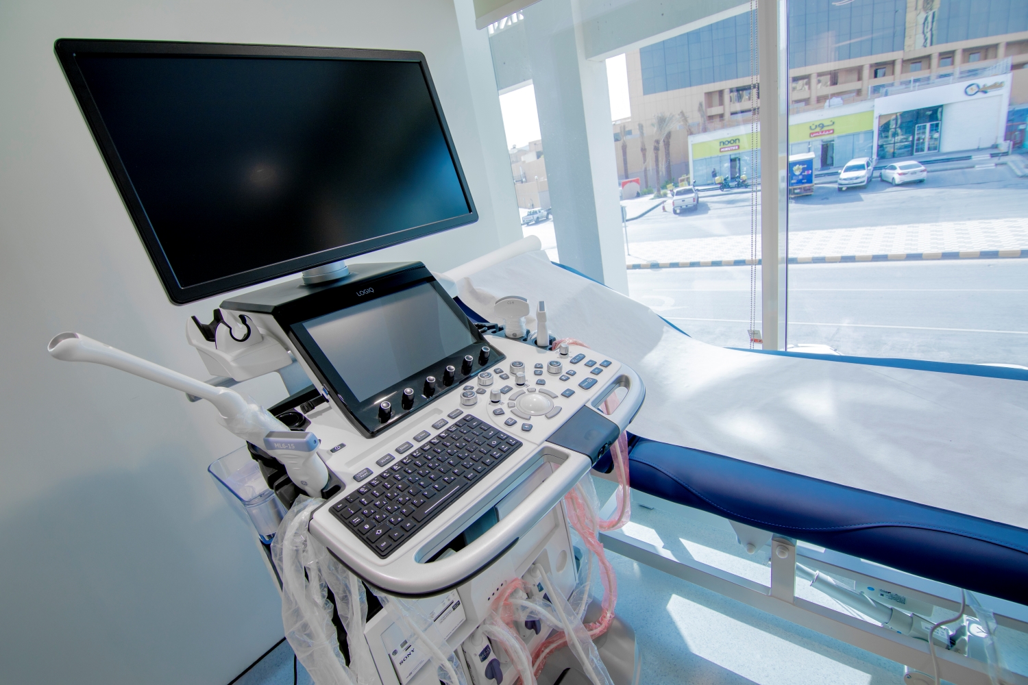
LOGIQ™ P9 Ultrasound & Doppler – Rabwah Branch Manufacturer: GE HealthCare
The LOGIQ™ P9 is a versatile ultrasound system designed for fast and accurate diagnostics across multiple clinical disciplines. It features advanced imaging technologies and a user-friendly interface.
Key Features:
1. Advanced Imaging Technologies:
- 2D Shear Wave Elastography: Quantitatively assesses tissue stiffness, aiding in the diagnosis and monitoring of chronic liver disease, breast conditions, etc.
- UGAP (Ultrasound-Guided Attenuation Parameter): Measures liver fat content to evaluate non-alcoholic fatty liver disease (NAFLD).
- B-Flow™ & B-Flow Color: Display blood flow without using Doppler, offering direct visualization of vessels with minimal artifacts.
- Contrast Imaging: Enhances image contrast and depth for detailed visualization of tissues and vessels.
- HD Color: High sensitivity for imaging small vessels and low-flow areas, boosting diagnostic confidence.
2. Smart User Interface:
- Scan Assistant: Step-by-step guide that reduces scan time.
- Auto Measure: AI-powered automatic measurements for precision and consistency.
- Compare Assistant: Facilitates side-by-side comparison with prior studies for better follow-up.
Clinical Applications:
- Abdominal Imaging: Liver, kidney, and intestinal disorders
- Obstetrics & Gynecology: Pregnancy, ovaries, uterus
- Cardiology: Heart and blood vessel imaging
- Urology: Bladder and kidney evaluation
- Soft Tissue: Muscles, tendons, glands
- Pediatrics: Imaging of tissues and organs in children
This system is ideal for healthcare providers seeking high-quality, advanced imaging with ease of use—enhancing diagnostic efficiency and overall patient care.
GE LOGIQ E10 Ultrasound & Doppler – Al-Namothajiyah & Al-Nafel Branches Manufacturer: GE HealthCare
The GE LOGIQ E10 is a high-performance ultrasound system delivering exceptional image quality, operational efficiency, and wide clinical support.
Key Features:
1. Superior Image Quality:
- Built with the cSound™ architecture, combining XDclear™ transducers, the cSound Imageformer processor, and Speckle Reduction Imaging—delivering crystal-clear images across various clinical scenarios.
2. Advanced Clinical Tools:
- 2D SWE (Shear Wave Elastography): Quantitative assessment of tissue stiffness for liver and other organ diagnostics.
- UGAP (Ultrasound-Guided Attenuation Parameter): Measures liver fat levels for fatty liver disease evaluation.
- CEUS (Contrast-Enhanced Ultrasound): Enhances soft tissue visualization and vascular assessment.
- Volume Navigation: Integrates ultrasound with other imaging modalities for precise interventional planning and guidance.
3. AI-Driven Support:
- OB Measurement Assistant & Auto Doppler Assistant: Streamlines exams and reduces scanning time.
- Hepatic Assistant: Combines 2D SWE and UGAP at the press of a button.
- Scan Assistant: Customizable protocols to improve workflow and standardization.
4. Data Security:
- Equipped with SonoDefense, a multi-layer cybersecurity system to protect patient data and ensure health information safety.
Clinical Applications:
- General Imaging: Abdomen, vascular, musculoskeletal
- Obstetrics & Gynecology: Fetal growth and maternal health monitoring
- Liver Imaging: Evaluation of fibrosis and fatty liver disease
- Cardiac Imaging: Heart structure and function assessment
- Interventional Imaging: Real-time guidance during minimally invasive procedures