Services
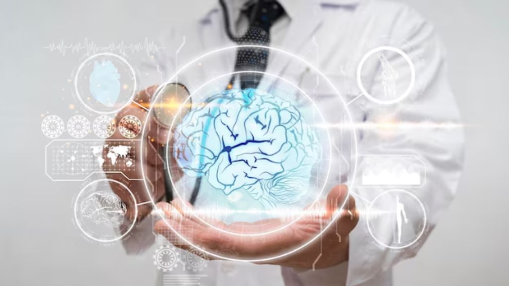
Neurological diagnostic unit
At RadCare Imaging Center, we provide a comprehensive range of advanced diagnostic methods to accurately assess the structure, functions, and diseases of the nervous system. Examinations are performed using non-invasive techniques, with the option of providing anesthesia/sedation when necessary, especially in MRI procedures to ensure patient comfort and stability.
First: Neuroimaging Techniques
1. Magnetic Resonance Imaging (MRI):
- Uses: Assessment of brain structure, tumors, multiple sclerosis (MS), strokes, and infections.
Advanced Studies:
- Functional MRI (fMRI): Mapping brain activity based on blood flow.
- Diffusion Tensor Imaging (DTI): Imaging of white matter pathways.
- Magnetic Resonance Spectroscopy (MRS): Measuring metabolic changes (e.g., tumors or metabolic diseases).
2. Computed Tomography (CT):
- Uses: Rapid assessment of brain hemorrhage, trauma, or hydrocephalus. Widely used in emergency cases.
3. Positron Emission Tomography (PET):
- Description: Tracing radioactive substances such as FDG or Amyloid to measure metabolic activity.
- Uses: Diagnosis of dementia (e.g., Alzheimer’s), tumors, and epileptic foci.
4. Angiography:
- Uses: Imaging blood vessels using non-invasive techniques such as CT/MR Angiography to evaluate arterial stenosis or occlusion.
5. Ultrasound:
Uses:
- Carotid Doppler: Assessing stenosis in neck arteries.
- Transcranial Doppler (TCD): Monitoring blood flow within the brain (e.g., vasospasm after subarachnoid hemorrhage).
Second: Electrical Activity Studies
1. Electroencephalography (EEG):
- Description: Recording brain electrical activity using scalp electrodes.
- Uses: Diagnosis of epilepsy, sleep disorders, and encephalopathy.
2. Electromyography and Nerve Conduction Studies (EMG & NCS):
- EMG: Measuring electrical activity in muscles – used for diagnosing conditions like Amyotrophic Lateral Sclerosis (ALS) or muscle diseases.
- NCS: Evaluating nerve conduction velocity – for diagnosing conditions such as neuropathy or Carpal Tunnel Syndrome.
3. Evoked Potentials (EP):
- Description: Recording electrical responses to sensory stimuli (visual, auditory, somatosensory).
- Uses: Assessment of multiple sclerosis, optic neuritis, and brainstem lesions.