Services
- Abdominal x-ray
- Abdominal Ultrasound
- Bone X-rays
- Cardiac MRI
- Carotid Doppler
- Chest X-Ray
- CT SCAN
- Echocardiography
- Electrocardiography (ECG)
- General Ultrasound Imaging
- General Doppler Imaging
- Magnetic Resonance Imagining
- Prostatic Ultrasound
- Scrotal Ultrasound
- Ultrasound of the Thyroid
- Venous Doppler
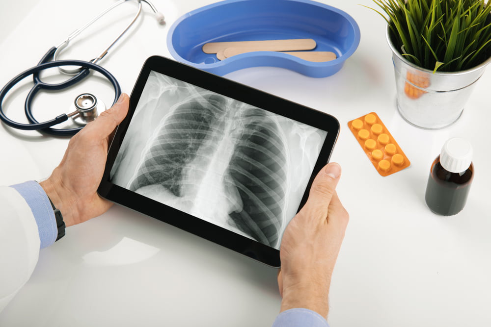
Professional medical service of Chest X-Ray
An X-ray, or radiograph, is a noninvasive diagnostic test that helps physicians diagnose and treat a variety of medical conditions. It works by exposing a part of the body to a small dose of ionizing radiation to create images of internal structures. X-rays are the oldest and most commonly used form of medical imaging.
Uses of a Chest X-Ray
A chest X-ray is performed to evaluate the lungs, heart, and chest wall. It is often the first imaging test used to investigate symptoms such as:
- Shortness of breath
- Persistent or severe cough
- Chest pain or injury
- Fever
Physicians use chest X-rays to diagnose or monitor treatment for conditions like:
- Pneumonia
- Heart failure and other heart-related issues
- Emphysema
- Lung cancer
- Line and tube placement
- Other medical conditions
Preparation Guidelines
- Most X-ray exams require no special preparation.
- You may be asked to remove some or all clothing during the exam.
- Remove any jewelry, removable dental appliances, eyeglasses, and other metal objects or clothing that might interfere with the imaging process.
- Women should always inform their physician and X-ray technologist if there is any possibility of pregnancy.