Professional medical service of Diagnostic Radiology Of Dental Imaging
What is it?
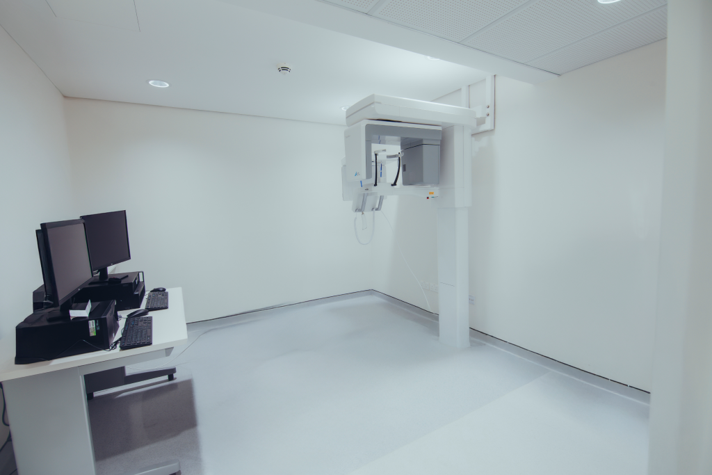
An x-ray (radiograph) is a noninvasive medical imaging test that helps physicians diagnose and treat various conditions. It works by exposing a part of the body to a small dose of ionizing radiation, creating detailed images of internal structures. X-rays are one of the oldest and most widely used forms of medical imaging.
Why is it Performed?
A panoramic x-ray is especially valuable for dental and oral health assessments because it:
- Covers a wider area than conventional intraoral x-rays.
- Provides critical information about:
- The nasal area
- Maxillary sinuses
- Tooth positioning
- Gum and bone irregularities
It is commonly used for planning:
- Dentures (full and partial)
- Braces
- Extractions
- Dental implants
Additional uses include detecting:
- Advanced periodontal disease
- Oral cysts
- Tumors and oral cancer
- Impacted teeth
- Temporomandibular joint disorder (TMJ)
- Sinusitis
Preparation Guidelines
- General Guidelines:
- Most x-ray exams require no special preparation.
- Clothing and Accessories:
- You may need to remove some or all clothing during the procedure.
- Remove any jewelry, removable dental appliances, eyeglasses, or metal objects that might interfere with the images.
- Pregnancy Precautions:
- Women should always inform their physician and x-ray technologist if there is any possibility of pregnancy.