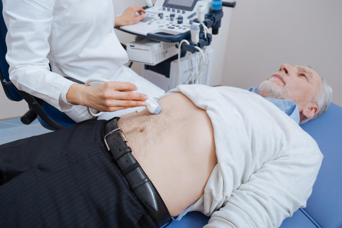
Professional medical service of Abdominal Ultrasound
Ultrasound imaging, also known as ultrasound scanning or sonography, is a safe and painless diagnostic technique that uses high-frequency sound waves to produce images of the body’s internal structures. A small probe and ultrasound gel are used to transmit sound waves, which create real-time images of internal organs and blood flow. Unlike X-rays, ultrasound does not use ionizing radiation.
Types of Ultrasound Imaging
- Conventional Ultrasound: Produces thin, flat images of the body.
- Doppler Ultrasound: Evaluates blood flow through major arteries and veins in areas like the abdomen, arms, legs, and neck.
Why Undergo an Abdominal Ultrasound?
Abdominal ultrasound evaluates the following organs and structures:
- Kidneys
- Liver
- Gallbladder
- Bile ducts
- Pancreas
- Spleen
- Abdominal aorta and other abdominal blood vessels
Conditions Diagnosed
Ultrasound helps diagnose a range of conditions, including:
- Abdominal pain or distention (enlargement)
- Abnormal liver function
- Enlarged abdominal organs
- Kidney stones
- Gallstones
- Abdominal aortic aneurysm
- Ultrasound is also used to guide biopsies and other medical procedures.
Preparation Guidelines
- Clothing: Wear loose, comfortable clothing. You may need to remove clothing or jewelry from the area being examined.
- Specific Preparations: Preparation depends on the type of examination:
- Liver, Gallbladder, Spleen, Pancreas: Eat a fat-free meal the evening before your test and avoid eating for 8–12 hours prior to the exam.
- Kidneys: Drink 4–6 glasses of liquid about an hour before the test to fill your bladder. Avoid eating for 8–12 hours beforehand to reduce intestinal gas.
- General: Your doctor may instruct you to avoid eating or drinking for up to 12 hours before your appointment.
- For Children: Ultrasound exams are sensitive to movement. For a smooth experience, explain the procedure to your child beforehand to help them remain calm and still during the test.